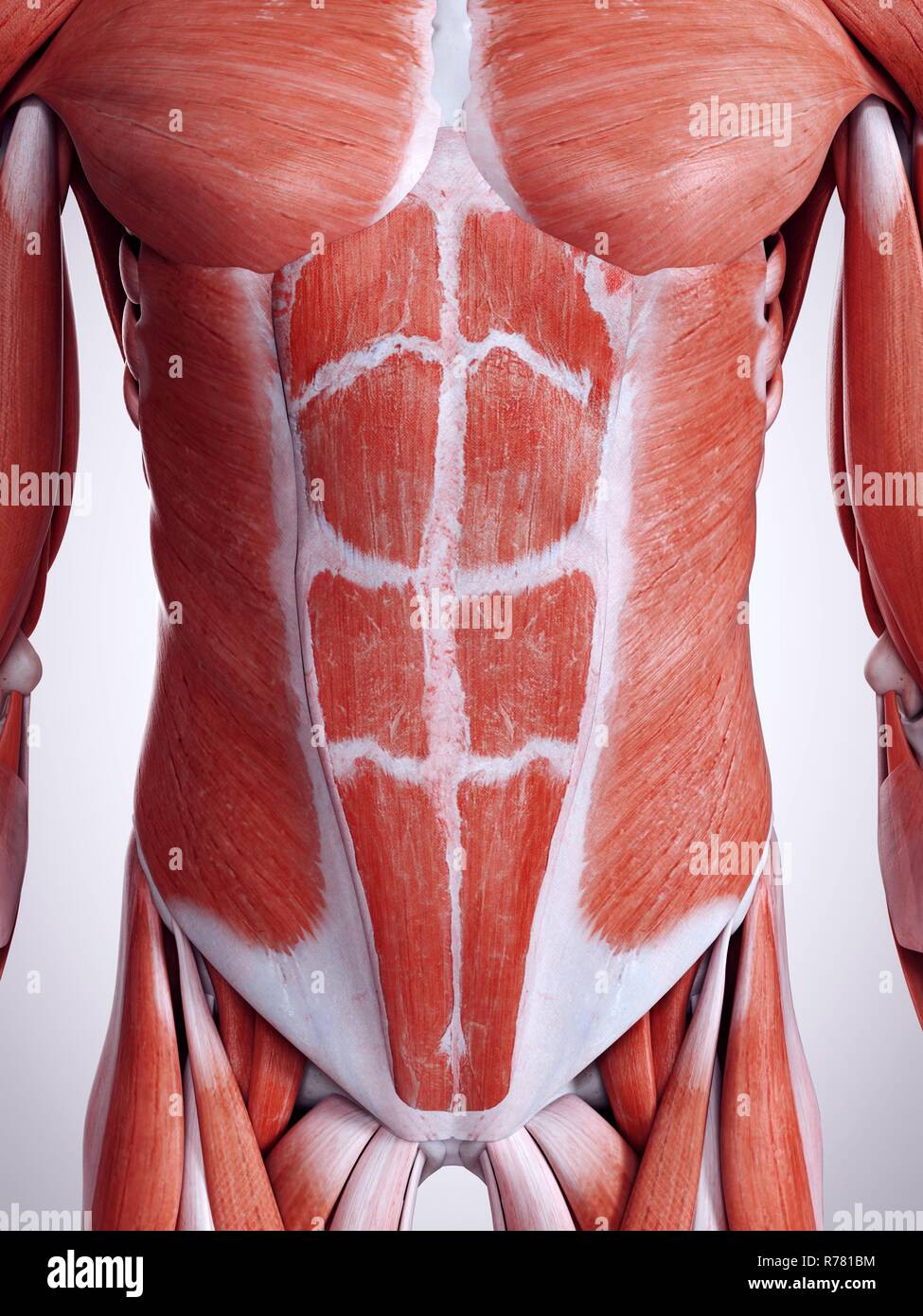
3d Illustration of the Internal Abdominal Muscles Anatomical Position

Complete Abdominal Organs 3D Model Collection CGTrader
Abdominal Organs - A 3D model collection by University of Dundee, CAHID (@anatomy_dundee)
Abdominal Anatomy Organs Instant Anatomy Abdomen Nerves
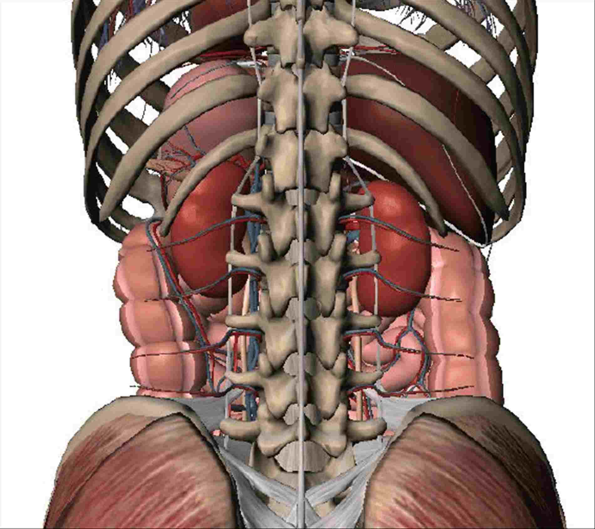
62 Download 3D Model Triangles: 324.9k Vertices: 165.1k More model information This model shows some of the organs and vessels in the abdomen. It's part of an E-Learning module about the inspection of the abdomen (Onderzoek van de buik). It is based on our previous model "Thorax and abdomen: some arteries and veins."
3D Model Update Ultrarealistic Abdomen & Spine regions unveiled in
Attached to the pelvis are muscles of the buttocks, the lower back, and the thighs. These muscles, including the gluteus maximus and the hamstrings, extend the thigh at the hip in support of the body's weight and propulsion. Other pelvic muscles, such as the psoas major and iliacus, serve as flexors of the trunk and thigh at the hip joint and.
Female abdominal organs, midsection, digital illustration. — biological
Abdomen. Home; 3D Human Body; Anatomy by Region; Abdomen; personAbdomen fullscreenclose. 3D model is a premium feature. Go Premium! Interactive 3D Models; Access over 1700 multiple choice questions; Advert Free; Custom Quiz Builder; Performance tracking; Upgrade to premium. Already have an account?
abdomen anatomy 3d
3D Model Update: Ultra-realistic Abdomen & Spine regions unveiled in summer release Posted on June 15, 2021 by 3D4Medical | 2 min read Creating a world class digital anatomy product is no small feat. To maintain this gold-standard in medical and anatomical education, we must stay on our toes and continually improve.

Abdominal anatomy, artwork Stock Image F006/0387 Science Photo
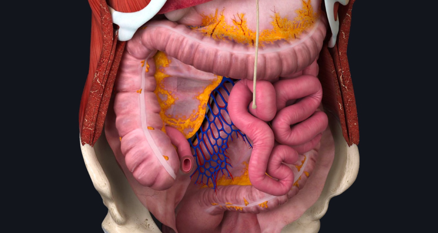
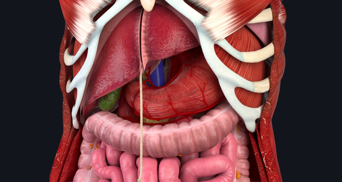
See this view in 3D! The lesser omentum connects the liver, stomach, and part of the duodenum. It helps hold the stomach in place and also serves as a support for blood and lymph vessels that supply the organs of the upper abdomen. Posterior view of the lesser omentum. Have VB Suite on your mobile device? See this view in 3D!
3d Abdomen Abdominal Anatomical Stock Photos & 3d Abdomen Abdominal
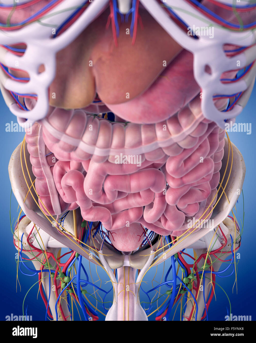
The abdomen is the body region found between the thorax and the pelvis. Its superior aperture faces towards the thorax, enclosed by the diaphragm. Inferiorly the abdomen is open to the pelvis, communicating through the superior pelvic aperture (pelvic inlet). These two apertures, together with abdominal walls, bound the abdominal cavity.

3d Rendered Image & Photo (Free Trial) Bigstock
The abdomen is the part of the body that contains all of the structures between the thorax (chest) and the pelvis, and is separated from the thorax via the diaphragm. The region occupied by the abdomen is called the abdominal cavity, and is enclosed by the abdominal muscles at front and to the sides, and by part of the vertebral column at the back.
📢 🥁 Introducing the new abdomen model available NOW in Complete Anatomy
You will explore the 3D anatomy of the organs from a basic level, providing thorough anatomical understanding, to its advanced application in surgical procedures. This course will challenge you to discover and help you to understand the anatomy of the abdomen and pelvis in all its aspects, ranging from its embryological underpinnings, via.
Normal Abdominal Organs Anatomy TrialExhibits Inc.
Introduction to the Digestive System Digestive System Basics - Mouth and Pharynx Digestive System Basics - Oesophagus and Stomach Digestive System Basics - Duodenum to Anus Digestive System Basics - Accessory Organs Peritoneal Cavity Intraperitoneal and Retroperitoneal Organs Ligaments/Peritoneal Attachments of the Liver Peritoneal Cavity
Male Abdominal Organs Photograph by Sebastian Kaulitzki/science Photo
3D anatomy tutorials and interactive modules on the muscles and layers of the anterior abdominal wall. Learn about the rectus abdominis and oblique muscles.
3d rendered illustration of the abdominal muscles Stock Photo Alamy
ANATOMY 3D ATLAS allows you to study human anatomy in an easy and interactive way. Through a simple and intuitive interface it is possible to observe, by highly detailed 3D models, every anatomical structure of the human body from any angle.
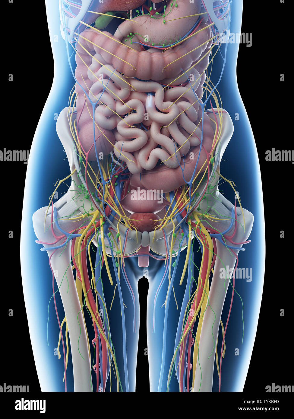
Ilustración 3D prestados de una anatomía abdominal hembras Fotografía
Human body Abdomen Abdomen The muscles of the abdomen protect vital organs underneath and provide structure for the spine. These muscles help the body bend at the waist. The major muscles of.

human abdominal organs 3d model
http://www.anatomyzone.com3D anatomy tutorial on the muscles of the abdominal wall using the Zygote Body Browser (http://www.zygotebody.com). This tutorial i.
3d rendered medically accurate illustration of the abdominal organs
Zygote Body is a free online 3D anatomy atlas. View, isolate, and learn human anatomy structures with Zygote Body. | |. Use the model select icon above the anatomy slider on the left to load different models. Premium Tools. My Scenes allows you to load and save scenes you have created. All annotations, pins and visible items will be saved.
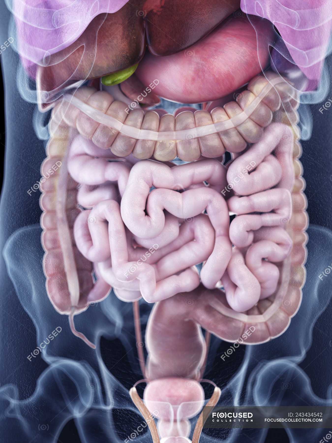
Illustration of human abdominal anatomy in body silhouette. — Digitally
Upon exiting the vertebral canal, the spinal nerves of the lower back form into two networks known as the lumbar and sacral plexuses. The lumbar plexus supplies nerves to the skin and muscles of the lateral abdominal region, thigh, anterior thigh, and external genitals. The sacral plexus similarly supplies nerves to the skin and muscles of the.
